3D Cone Beam Imaging Portland
Extreme Diagnostic Precision for Outstanding Results
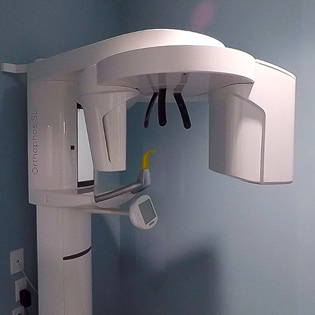
Three-dimensional (3D) cone beam imaging or cone beam computed tomography (CBCT) is a special type of X-ray equipment used when regular dental or facial X-rays are not sufficient. This advanced 3D imaging system is used to produce 3D images of your teeth, soft tissues, nerve pathways and bone in the craniofacial region in a single scan.
Images obtained with cone beam CT allow for more precise treatment planning.
How 3D Cone Beam Imaging Works
CBCT can be used to produce images that are similar to those produced by conventional CT imaging. With cone beam CT, an X-ray beam in the shape of a cone is moved around the patient to produce a large number of images, also called views. CT scans and cone beam CT both produce high-quality images.
Dental cone beam CT was developed as a means of producing similar types of images but with a much smaller and less expensive machine that could be placed in an outpatient office. Dental cone beam CT is commonly used for treatment planning of orthodontic issues. It is also useful for more complex cases that involve:
- Planning surgery for impacted teeth
- Diagnosing temporomandibular joint disorder (TMJ)
- Placing dental implants accurately
- Evaluating the jaw, sinuses, nerve canals and nasal cavity
- Detecting, measuring and treating jaw tumors
- Determining the bone structure and tooth orientation
- Locating the origin of pain or pathology
- Performing cephalometric analysis
- Planning reconstructive surgery
Benefits of 3D Cone Beam Imaging

Advanced 3D imaging provides patients with the most complete story of their oral health, and provides them the ability to make the best decisions with less hesitation.
Additionally, advanced 3D imaging methods do not expose you to as much radiation as traditional imaging methods, because traditional imaging methods take a longer amount of time (5-10 minutes) to complete and you have to be exposed multiple times to get a complete picture. Advanced dental imaging takes a fraction of the time (10 seconds) so you are exposed to less harmful radiation.
Cone Beam CT vs. Conventional CT

Dental cone beam CT provides detailed images of the bone and is performed to evaluate diseases of the jaw, dentition, bony structures of the face, nasal cavity, and sinuses. It does not provide the full diagnostic information available with conventional CT, particularly in the evaluation of soft tissue structures such as muscles, lymph nodes, glands, and nerves. However, cone beam CT has the advantage of lower radiation exposure compared to conventional CT.
What to Expect When Getting 3D Cone Beam Imaging
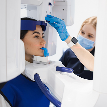
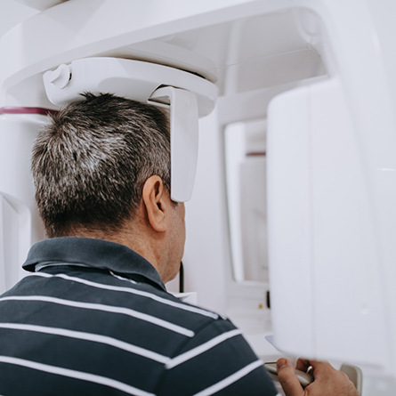
You will be asked to bite on a jaw positioner and hold it. Once the area of interest is centered in the beam of the machine, you will be asked to remain very still while the X-ray source and detector revolve around you for a 360-degree rotation or less. This typically can take between 20 to 40 seconds, during which the machine captures multiple images from different angles that are then reconstructed to create a single 3D image of your mouth.
The X-ray source and detector are mounted on opposite sides of the revolving C-arm or gantry and rotate in unison. In a single rotation, the detector can generate anywhere between 150 to 200 high resolution two-dimensional (2D) images, which are then digitally combined to form a 3D image that can provide your dentist or oral surgeon with valuable information about your oral and craniofacial health.
Who Interprets the Cone Beam Imaging Results and How Do You Get Them?

Your dentist will analyze the images and discuss the results with you directly or communicate the results to your referring physician or dentist. With 3D digital imaging, the results are detailed and can be enhanced with digital imaging software for region-of-interest (ROI) magnification and equalization.
Why Choose a Dental Office with 3D Cone Beam Imaging?

At Belmont Family Dentistry, we invest in the latest technology, so we can offer our patients exceptional patient care. Our advanced 3D imaging takes a panoramic view of your mouth. This level of detail helps us improve our diagnostic accuracy which means you receive the best possible dental care.
Advanced 3D imaging also provides a detailed full picture of the mouth and skull from every angle providing more information than what a regular x-ray can provide. This is an excellent source for planning surgery.
3D Cone Beam Imaging FAQs
How Should You Prepare for a Dental Cone Beam CT?
This procedure requires little to no special preparation. Tell your dentist if there’s a possibility you are pregnant.
Remove any metal objects prior to the examination, including jewelry, piercings (if possible), eyeglasses, dentures or other removable dental work, hearing aids, and hairpins, as they may interfere with the imaging.
What Will You Experience During and After the Dental Cone Beam Procedure?
You will not experience any pain during a cone beam CT exam, and you will be able to return to your normal activities once the exam is complete.
Are 3D Dental X-Rays Safe?
The radiation emitted from a 3D X-ray machine is extremely small. If you are worried about this topic, we are happy to discuss your concerns at your appointment.